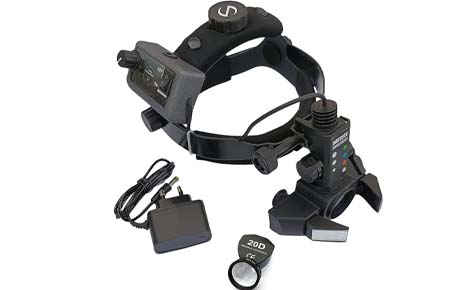
The 20D ophthalmoscope lens is a specialized, hand-held condensing lens used in combination with a binocular indirect ophthalmoscope (BIO) to perform a comprehensive examination of the posterior segment (fundus) of the eye. It is a gold standard tool in ophthalmology and optometry due to its excellent balance of magnification and field of view.
20D Ophthalmoscope
Overview
Key Features and Specifications
- Purpose: The primary function is for general and detailed examination of the retina, optic nerve head, and macula. It is also used for diagnosing and managing conditions like diabetic retinopathy, retinal detachments, and macular degeneration.
- Magnification: It provides an approximate 3.13x (or ~3x) magnification of the retina, which offers a detailed view of the structures.
- Field of View: The lens typically offers a wide field of view, ranging from 46 to 60 degrees, allowing visualization up to the mid-peripheral retina in a single image.
- Working Distance: It features a comfortable working distance of around 50 mm (5 cm) from the patient's eye, which allows for easy manipulation by the examiner.
- Image Orientation: When used with a BIO, the 20D lens produces a real, inverted (both horizontally and vertically) image of the fundus.
- Optics: Most modern 20D lenses use a high-quality double aspheric design, which minimizes distortions and ensures superior image clarity and color fidelity.
Clinical Use
The 20D lens is an incredibly versatile tool.
- Routine Exams: It is widely used in routine adult eye exams for general diagnosis.
- Pediatric Exams: Higher-powered lenses are sometimes preferred for small children or patients with small pupils, but the 20D remains a staple, particularly for training students and residents.
- Surgical Assessment: Ophthalmic surgeons use the lens to assess the retina before and after procedures like vitrectomy or retinal detachment repair.
- Dynamic Examination: The wide field of view allows for dynamic examination; the patient can look in various directions to help the practitioner visualize the far peripheral retina, especially when used with scleral depression.
- Smartphone Integration: The lens can even be combined with a smartphone camera to capture digital images or video of the fundus for documentation, teaching, or telemedicine purposes.