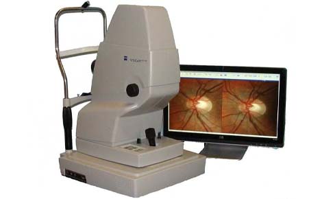
A fundus camera is a specialized low-power microscope with a camera, used to capture detailed images of the eye's interior (fundus) – retina, optic nerve, macula, blood vessels – for diagnosing vision-threatening conditions like diabetic retinopathy, glaucoma, macular degeneration, and tumors, using light and complex optics similar to an indirect ophthalmoscope. Modern versions include handheld, wide-angle, and AI-enhanced models, allowing non-invasive screening and monitoring, even without dilating drops (non-mydriatic).
Fundus Camera
Overview
Key Components & Features
- Microscope & Camera: Combines magnification with digital imaging.
- Optics: Uses lenses, mirrors, and beam splitters to direct light and capture images.
- Angle of View: Standard (around 30°), wide-angle (45°-140°), or narrow-angle (20° or less).
- Non-Mydriatic: Some cameras work without pupil-dilating drops, making exams quicker.
- Special Filters: Can be used for angiography (Fluorescein, ICG) or autofluorescence.