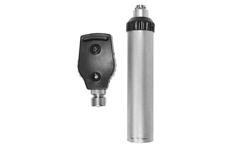
A direct ophthalmoscope is a portable, handheld tool for examining the eye's interior (fundus) by projecting light and providing a magnified, upright, virtual image of the retina, optic disc, and vessels, ideal for preliminary checks in primary care due to its portability and ability to work with undilated pupils, though it offers a limited, monocular view compared to indirect methods. It uses a system of mirrors, lenses, and a light source, allowing clinicians to assess overall eye health, detect issues like retinal detachments or cataracts, and check the optic disc's cup-to-disc ratio.
Direct Ophthalmoscope
Overview
How it Works
- Light & View: A light source shines through a mirror at a 45-degree angle into the patient's eye, illuminating the fundus.
- Image Formation: Light reflects back through the mirror and a lens system, forming a magnified, upright, virtual image that the examiner sees through the device.
- Magnification: Offers up to 15x magnification, the highest of fundus examination techniques, but with a narrow field of view.
Key Features & Use
- Portability: Small, handheld, and great for quick assessments.
- Examination: Allows viewing of the retina, optic disc, macula, and vessels.
- Patient Positioning: Examiner holds the device close to their eye and the patient's eye, with both potentially sitting or standing.
- Lens System: Rotating lenses help focus on structures and compensate for refractive errors, noted by the clarity of the red reflex.
- Anterior Segment: Can also provide a gross view of the anterior structures with added lenses.