A surgical microscope is a high-precision optical instrument used in operating rooms for magnified, illuminated, stereoscopic views of tiny structures, enabling delicate microsurgery in fields like neurosurgery, ophthalmology, ENT, and vascular repair, featuring adjustable zoom, focus, coaxial lighting, and often integrated with advanced imaging (OCT, AR) for enhanced precision and accuracy, controlled by a foot pedal for hands-free operation.
Surgical Microscope
Overview
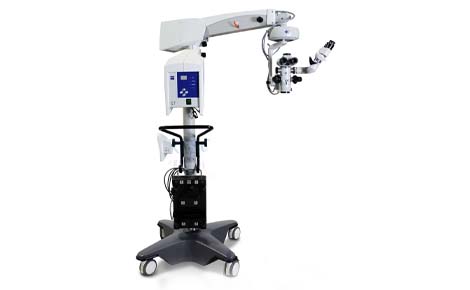
Key Components & Functionality
- Optics: Binocular eyepieces provide stereoscopic (3D) vision, while telescopic magnifiers offer zoom, ensuring clear, magnified views.
- Illumination: Usually coaxial (following the light path to avoid shadows) and fiber-optic, with sources like Xenon or LED, keeping heat away from the surgical field.
- Mounting: A heavy floor stand with wheels and a suspension arm allows precise positioning and stabilization, with brakes to lock it in place.
- Control: A foot pedal controls focus, zoom, XY positioning, and illumination, freeing the surgeon's hands.
Key Applications
- Ophthalmology: Cataract, retinal, and corneal surgeries (keratoplasty).
- Neurosurgery: Brain tumor, aneurysm, and spinal surgeries.
- ENT (Ear, Nose, Throat): Procedures like tympanostomy tube insertion.
- Vascular Surgery: Micro-anastomosis (joining blood vessels).
- Plastic/Reconstructive Surgery: Delicate tissue repair.