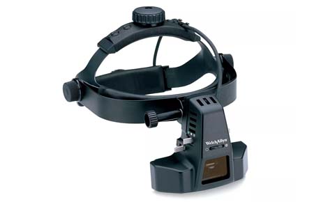
An indirect ophthalmoscope is a head-worn device with a light and eyepieces, used with a handheld high-plus condensing lens to view the back of the eye (fundus). It offers a wide, stereoscopic (3D) view of the retina with 2-5x magnification, excellent illumination (even through cataracts), and is vital for diagnosing retinal tears, detachments, and systemic disease effects, requiring dilated pupils for best results. Examiners see a real, inverted image of the fundus, requiring practice to interpret, but allowing detailed peripheral retinal exams via scleral indentation.
Indirect Ophthalmoscope
Overview
How it works
- Illumination: A bright light from the scope enters the patient's eye.
- Condensing Lens: The examiner holds a powerful convex lens (e.g., +20D) near the eye.
- Image Formation: Light from the retina passes through the lens, which converges it to form a real, inverted, magnified image of the fundus in the space between the lens and the examiner.
- Viewing: The examiner looks through the binocular scope to see this inverted image, getting a 3D view.
Key Features & Benefits
- Wide Field of View: Examines a larger area of the retina than direct ophthalmoscopy.
- Stereoscopic (3D) Vision: Binocular version provides depth perception.
- Superior Illumination: Bright light helps see through cloudy media like cataracts.
- Peripheral Access: Allows use of scleral indentation to view the far periphery.
- Diagnostic Power: Essential for retinal tears, detachment, diabetes, hypertension.